
Petrophysical characterization of Indiana limestone using medical dual-energy computed tomography technique: insights into porosity, bulk density, and effective atomic number
DOI:
https://doi.org/10.69631/s3tb8r43Keywords:
Digital Rock Physics, Petrophysics, Dual-energy CT technique, DECT, Medical CTAbstract
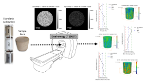
Computed Tomography (CT) enables non-destructive 3D reconstruction of pore structures and rock properties mapping. Typically, such images are obtained from micro-CT (µm-scale) or synchrotron imaging (nm-scale). Despite their accuracy, these high-resolution imaging methods are expensive, time-consuming, and limited in sample size, affecting representative volume analysis. This work investigates an alternative approach using medical-CT (100 µm resolution), applying the dual-energy CT technique (DECT) to characterize petrophysical properties (total porosity, bulk density, and effective atomic number) of six Indiana limestone samples (3.81 cm diameter, 4.88 cm length). Samples with porosities between 17.5% and 19.1% were scanned using paired high-energy (130 kV) and low-energy (80 kV) protocols, allowing DECT analysis to generate detailed 2D and 3D property maps. Additionally, the single-energy CT (SECT) technique, enhanced by subtracting images of the rock sample in saturated and dry conditions, improved the estimation of effective porosity. The results from DECT and SECT, processed with Python scripts and Avizo 3D, demonstrated average differences of 3.34% for bulk density, 5.30% for effective porosity, and 4.65% for effective atomic number compared to basic petrophysics measurements. Although artifacts from low-energy scans presented limitations, their impact can be reduced by optimizing acquisition parameters, improving the experimental setup, and applying reconstruction filtering techniques. Overall, this study highlights medical-CT as a fast, cost-effective method for analyzing larger samples, providing a practical alternative to traditional high-resolution imaging to estimate key petrophysical properties for the identification and visualization of heterogeneity on carbonates rocks.
Downloads
References
1. Akin, S., & Kovscek, A. R. (2003). Computed tomography in petroleum engineering research. Geological Society, London, Special Publications, 215(1), 23–38. https://doi.org/10.1144/GSL.SP.2003.215.01.03
2. Antelo, W. L. F., Vargas, V., & Moreno, R. B. Z. L. (2025). Multiscale reconstruction of carbonate rock pore structure using medical and micro-ct imaging [conference poster]. https://doi.org/10.13140/RG.2.2.15904.85761
3. Archilha, N. L., Costa, G. R., Ferreira, G. R. B., Moreno, G. B. Z. L., Rocha, A. S., Meyer, B. C., Pinto, A. C., Miqueles, E. X. S., Cardoso, M. B., & Westfahl, H. (2022). Mogno, the nano and microtomography beamline at Sirius, the Brazilian synchrotron light source. Journal of Physics: Conference Series. Institute of Physics, 2380(1), Article 012123. https://doi.org/10.1088/1742-6596/2380/1/012123
4. Bazaikin, Y., Gurevich, B., Iglauer, S., Khachkova, T., Kolyukhin, D., Lebedev, M., Lisitsa, V., & Reshetova, G. (2017). Effect of ct image size and resolution on the accuracy of rock property estimates. Journal of Geophysical Research: Solid Earth, 122(5), 3635–3647. https://doi.org/10.1002/2016JB013575
5. Bhattad, P., Young, B., Berg, C. F., Rustad, A. B., & Lopez, O. (September 2014). X-ray micro-ct assisted drainage rock typing for characterization of flow behaviour of laminated sandstone reservoirs. In Proceedings of the international symposium of the Society of Core Analysts (SCA), Avignon, France. http://refhub.elsevier.com/S0309-1708(16)30309-8/sbref0005
6. Brabant, L., Dierick, M., Pauwels, E., De Witte, Y., & Van Hoorebeke, L. (2013). Modifications of iterative reconstruction algorithms for the reduction of artefacts in high resolution x-ray computed tomography, 7, 111–114.
7. Buades, A., Coll, B., & Morel, J.-M. (2005). A non-local algorithm for image denoising. In IEEE Computer Society Conference on Computer Vision and Pattern Recognition (CVPR’05), 2 (pp. 60–65). https://doi.org/10.1109/CVPR.2005.38
8. Bultreys, T., De Boever, W., & Cnudde, V. (2016). Imaging and image-based fluid transport modeling at the pore scale in geological materials: A practical introduction to the current state-of-the-art. Earth-Science Reviews, 155, 93–128. https://doi.org/10.1016/j.earscirev.2016.02.001
9. Chaves, J. M. P., & Moreno, R. B. Z. (2021). Low- and high-resolution x-ray tomography helping on petrophysics and flow-behavior modeling. SPE Journal, 26(1), 206–219. https://doi.org/10.2118/202495-PA
10. Chen, B., Duan, X., Yu, Z., Leng, S., Yu, L., & McCollough, C. [Technical note]. (2015). Technical Note: Development and validation of an open data format for ct projection data. Medical Physics, 42(12), 6964–6972. https://doi.org/10.1118/1.4935406
11. du Plessis, A., Le Roux, S. G., & Guelpa, A. (2016). Comparison of medical and industrial x-ray computed tomography for non-destructive testing. Case Studies in Nondestructive Testing and Evaluation, 6, 17–25. https://doi.org/10.1016/j.csndt.2016.07.001
12. du Plessis, A., & Rossouw, P. (2015). X-ray computed tomography of a titanium aerospace investment casting. Case Studies in Nondestructive Testing and Evaluation, 3, 21–26. https://doi.org/10.1016/j.csndt.2015.03.001
13. Erol, S., Fowler, S. J., Harcouët-Menou, V., & Laenen, B. (2017). An analytical model of porosity–permeability for porous and fractured media. Transport in Porous Media, 120(2), 327–358. https://doi.org/10.1007/s11242-017-0923-z
14. Fitzhenry, E., Martel, R., Robert, T., & Roches, M. D. (2022). Dual-energy ct scan protocol optimization to monitor transient fluid saturation distributions during three-phase flow in sand columns. Colloids and Surfaces. Part A: Physicochemical and Engineering Aspects, 645(7), Article 128955. https://doi.org/10.1016/j.colsurfa.2022.128955
15. Freire-Gormaly, M., Ellis, J. S., MacLean, H. L., & Bazylak, A. (2015). Pore structure characterization of indiana limestone and pink dolomite from pore network reconstructions. Oil & Gas Science and Technology—Revue d’IFP Energies Nouvelles, 71(3), 33. https://doi.org/10.2516/ogst/2015004
16. Gamal, H., Bageri, B. S., Elkatatny, S., & Patil, S. (2021). Investigating the alteration of sandstone pore system and rock features by role of weighting materials. ACS Omega, 6(5), 4100–4110. https://doi.org/10.1021/acsomega.0c06256
17. Garnier, P., Angulo-Jaramillo, R., Dicarlo, D. A., Bauters, T. W. J., Darnault, C. J. G., Steenhis, T. S., Parlange, J.-Y., & Baveye, P. (1998). Dual-energy synchrotron x ray measurements of rapid soil density and water content changes in swelling soils during infiltration. Water Resources Research, 34(11), 2837–2842. https://doi.org/10.1029/98WR02367
18. Ketcham, R. A., & Carlson, W. D. (2001). Acquisition, optimization and interpretation of x-ray computed tomographic imagery: Applications to the geosciences. Computers and Geosciences, 27(4), 381–400. https://doi.org/10.1016/S0098-3004(00)00116-3
19. Korneev, S. V., Yang, X., Zachara, J. M., Scheibe, T. D., & Battiato, I. (2018). Downscaling-based segmentation for unresolved images of highly heterogeneous granular porous samples. Water Resources Research, 54(4), 2871–2890. https://doi.org/10.1002/2018WR022886
20. Lin, Q., Al-Khulaifi, Y., Blunt, M. J., & Bijeljic, B. (2016). Quantification of sub-resolution porosity in carbonate rocks by applying high-salinity contrast brine using x-ray microtomography differential imaging. Advances in Water Resources, 96, 306–322. https://doi.org/10.1016/j.advwatres.2016.08.002
21. Madonna, C., Quintal, B., Frehner, M., Almqvist, B. S. G., Tisato, N., Pistone, M., Marone, F., & Saenger, E. H. (2013). Synchrotron-based x-ray tomographic microscopy for rock physics investigations. Geophysics, 78(1), D53–D64. https://doi.org/10.1190/GEO2012-0113.1
22. Mendieta-Penagos, A., Rincón-Bautista, L. R., & Herrera-Otero, E. (2019). Español [Quantitative determination of heterogeneity in samples of rock type plug using x-ray computerized tomography]. Boletin de Geologia, 41(1), 133–149. https://doi.org/10.18273/revbol.v41n1-2019007
23. Moreira, A. C., Santos, V. A., Mantovani, I. F., Cunha Neto, J. A. B., & Fernandes, C. P. (2021). Evaluation of the induced oil remobilization through high and low salinity waterflooding in a porous system via x-ray microtomography. Chemical Engineering Transactions, 86, 1159–1164. https://doi.org/10.3303/CET2186194
24. Moslemipour, A., & Sadeghnejad, S. (2021). Dual-scale pore network reconstruction of vugular carbonates using multi-scale imaging techniques. Advances in Water Resources, 147(1), Article 103795. https://doi.org/10.1016/j.advwatres.2020.103795
25. Pini, R., & Madonna, C. (2016). Moving across scales: A quantitative assessment of x-ray ct to measure the porosity of rocks. Journal of Porous Materials, 23(2), 325–338. https://doi.org/10.1007/s10934-015-0085-8
26. Ramstad, T., Idowu, N., Nardi, C., & Øren, P. E. (2012). Relative permeability calculations from two-phase flow simulations directly on digital images of porous rocks. Transport in Porous Media, 94(2), 487–504. https://doi.org/10.1007/s11242-011-9877-8
27. Ramstad, T., Øren, P.-E., & Bakke, S. (2009). Simulation of two phase flow in reservoir rocks using a lattice Boltzmann method. In SPE annual technical conference and exhibition. SPE. https://doi.org/10.2118/124617-MS
28. Roberto Carvalho de Holleben, C. R. (1993). Determinação de porosidade e saturações de fluidos atraves da tomografia computadorizada de raios-x. Handle.Net (pp. 1–198). https://hdl.handle.net/20.500.12733/1581369. https://doi.org/10.47749/T/UNICAMP.1993.75955
29. Ruspini, L. C., Øren, P. E., Berg, S., Masalmeh, S., Bultreys, T., Taberner, C., Sorop, T., Marcelis, F., Appel, M., Freeman, J., & Wilson, O. B. (2021). Multiscale digital rock analysis for complex rocks. Transport in Porous Media, 139(2), 301–325. https://doi.org/10.1007/s11242-021-01667-2
30. Shi, H., Yang, Z., & Luo, S. (2017). Reduce beam hardening artifacts of polychromatic x-ray computed tomography by an iterative approximation approach. Journal of X-Ray Science and Technology, 25(3), 417–428. https://doi.org/10.3233/XST-16187
31. Siddiqui, S. (2023). Some useful guidelines for whole core ct-scanning for petrophysical applications. E3S Web of Conferences. EDP Sciences, 367, Article 01013. https://doi.org/10.1051/e3sconf/202336701013
32. Siddiqui, S., & Khamees, A. A. (2004). Dual-energy CT-scanning applications in rock characterization. In SPE annual technical conference and exhibition. SPE. https://doi.org/10.2118/90520-MS
33. Siddiqui, S., Nasr-El-Din, H. A., & Khamees, A. A. (2006). Wormhole initiation and propagation of emulsified acid in carbonate cores using computerized tomography. Journal of Petroleum Science and Engineering, 54(3–4), 93–111. https://doi.org/10.1016/j.petrol.2006.08.005
34. Singh, B., Agrawal, A. K., Kashyap, Y. S., & Gadkari, S. C. (2017). X-ray synchrotron dual energy imaging for material specific study (Article 060010). Author(s). https://doi.org/10.1063/1.4980415
35. Soares, J. A., Garcia, A. J. V., Bezerra, F. H. R., Barbosa, J. A., Friedrich, A., Cazarin, C. L., Tabosa, L. D. G., & Coura, R. L. C. (2015). Petrophysics and rockphysics of carbonates from Brazil and Portugal. In 14th International Congress of the Brazilian Geophysical Society & EXPOGEF, Rio de Janeiro, Brazil, 3-6 August 2015 (pp. 882–887). Brazilian Geophysical Society. https://doi.org/10.1190/sbgf2015-173
36. Sousa, L., Menningen, J., López-Doncel, R., & Siegesmund, S. (2021). Petrophysical properties of limestones: Influence on behaviour under different environmental conditions and applications. Environmental Earth Sciences, 80(24). https://doi.org/10.1007/s12665-021-10064-3
37. Spurin, C., Ellman, S., Sherburn, D., Bultreys, T., & Tchelepi, H. A. (2024). Python workflow for segmenting multiphase flow in porous rocks. Transport in Porous Media, 151(15), 2819–2834. https://doi.org/10.1007/s11242-024-02136-2
38. Sulieman, H., Jouini, M. S., Alsuwaidi, M., Al-Shalabi, E. W., & Al Jallad, O. A. (2024). Multiscale investigation of pore structure heterogeneity in carbonate rocks using digital imaging and scal measurements: A case study from Upper Jurassic limestones, abu dhabi, uae. PLOS One, 19(2), Article e0295192. https://doi.org/10.1371/journal.pone.0295192
39. Surmas, R., & Ramalho Albuquerque, M. (2024). Synergies between laboratory and digital rock for improvement of rock models. In SPWLA 65th Annual Symposium transactions. Society of Petrophysicists and Well Log Analysts. https://doi.org/10.30632/SPWLA-2024-0135
40. Teles, A. P., Lima, I., & Lopes, R. T. (2016). Rock porosity quantification by dual-energy x-ray computed microtomography. Micron, 83, 72–78. https://doi.org/10.1016/j.micron.2016.02.004
41. Thermo Fisher Scientific. (2024). Amira-Avizo software 2024.1 user guide. Thermo Fisher Scientific, Incorporated. https://www.thermofisher.com/amira-avizo
42. Tino, R., Yeo, A., Brandt, M., Leary, M., & Kron, T. (2021). The interlace deposition method of bone equivalent material extrusion 3d printing for imaging in radiotherapy. Materials and Design, 199(2), Article 109439. https://doi.org/10.1016/j.matdes.2020.109439
43. Vargas, V., Antelo, W. L. F., & Moreno, R. B. Z. L. (2025). Characterization of sandstone and carbonate rocks using dual-energy x-ray computed tomography for sample selection in coreflooding tests. https://doi.org/10.5281/zenodo.16815648
44. Vinegar, H. J., & Wellington, S. L. (1987). Tomographic imaging of three-phase flow experiments. Review of Scientific Instruments, 58(1), 96–107. https://doi.org/10.1063/1.1139522
45. Wellington, S. L., & Vinegar, H. J. (1987). X-ray computerized tomography. Journal of Petroleum Technology, 39(8), 885–898. https://doi.org/10.2118/16983-PA
46. Wennberg, O. P., & Rennan, L. (2018). A brief introduction to the use of X-ray computed tomography (CT) for analysis of natural deformation structures in reservoir rocks. Geological Society, London, Special Publications, 459(1), 101–120. https://doi.org/10.1144/SP459.10
47. Withjack, E. M., Devier, C., & Michael, G. (2003). The role of X-ray computed tomography in core analysis. In SPE western regional/AAPG pacific section joint meeting. SPE. https://doi.org/10.2118/83467-MS
Downloads
Published
Data Availability Statement
All data generated and analyzed during this study, including CT images, properties maps, and python scripts, are available at https://redu.unicamp.br/dataset.xhtml?persistentId=doi:10.25824/redu/ZQOI8E.
Issue
Section
License
Copyright (c) 2026 Walter Leonardo Flores Antelo, Janeth Alina Vidal Vargas, Rosangela Barros Zanoni Lopes Moreno

This work is licensed under a Creative Commons Attribution-NonCommercial-NoDerivatives 4.0 International License.
This article is published under the Creative Commons license indicated above. See the license link for details.
Article metadata are available under the CCo license.
How to Cite
Funding data
-
Equinor
Grant numbers Project ANP 23271-0


